Xpolar® technology is an imaging mode for measuring
the birefringence of the samples.
The birefringence value is measured in each pixel of the image.

Figure 1 : Diagram of the XPolar® microscope and the interpretation of Kmax images
The XPolar® measurement
Electromagnetic Waves
Light can be thought of as an electromagnetic (EM) wave,
an example of this wave is shown in Fig. 2.
The polarization of the light [1] represents the direction of the oscillation
of its electric field (in red): The polarization of the electromagnetic wave
in Fig. 2 is vertical linear.
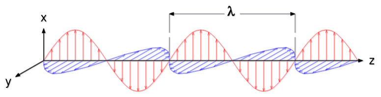
Figure 2 : Electromagnetic wave propagate along the z axis.
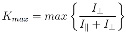
The intensity at illumination is polarized according to a given state of polarization S1.
Two intensities are measured : I∥ (resp. I⊥) by projecting the detected intensity
according to the polarization state parallel (resp. perpendicular) to S1.
Polarization state
|
The state of polarization of a wave represents |
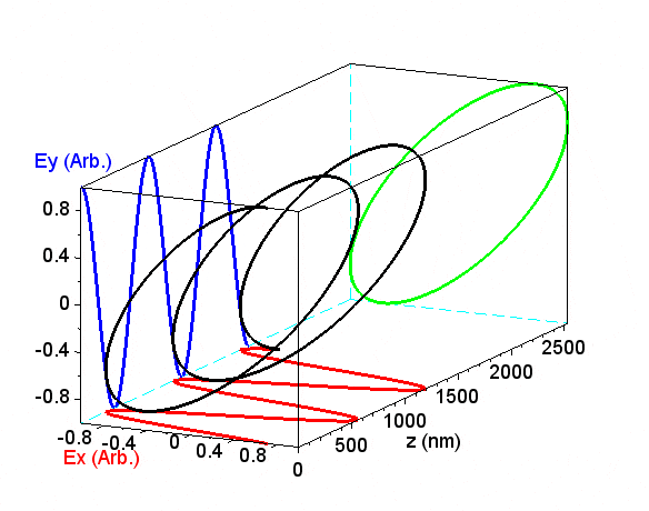
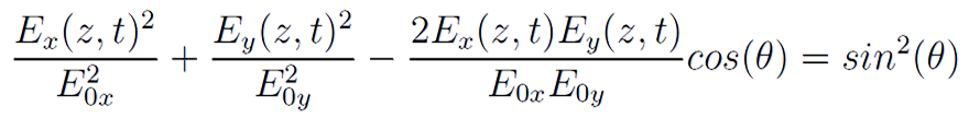
With : Ex(z, t) (resp. Ey(z, t)) the sinusoidal amplitude of the electric field projected on the x axis (resp. y),
E0x (resp. E0y) the amplitude at the origin on the x axis (resp. y),
θ the value of the phase shift between the two sinusoids Ex(z, t) et Ey(z, t).
Birefringent materials modify the value of the phase difference between the two sinusoids Ex(z, t) et Ey(z, t).
The set of polarization states can be represented on the Poincaré sphere.
XPolar® technology makes it possible to probe any state in the Poincaré sphere, with a
fixed frame of reference, i.e. without having to change the orientation of the sample or microscope.
XPolar® technology gives a measure of the value of Kmax for each pixel in the field of vision.
Kmax
The value of Kmax< is then obtained with :
This value is related to the birefringence of
the sample via relationship :

with θ the phase shift produced by birefringence Δn
of the sample thickness e, at wavelength λ.
Application : Skin, Collagen Fibers
Collagens are part of structural proteins.
Present in the extracellular matrix of certain living organisms,
they give the fabrics mechanical resistance to stretching.
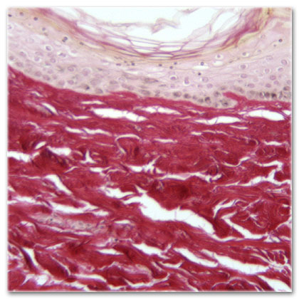
They are therefore present in large quantities in the skin (Fig. 3a)
 |
 |
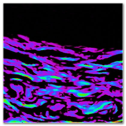 |
Figure 3a : Brightfield microscopy |
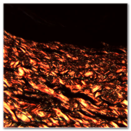
Figure 3b : Cross polarizerss |
Figure 3c : XPolar® technology |
They also have the property of modifying the polarization state of light,
intrinsically or after the addition of a specific dye. This property can be observed qualitatively
between crossed polarizers (Fig. 3b, the collagen appearing yellow / red), but this does not allow
to quantify any changes in the state of collagen [2].
XPolar® technology makes it possible to quantify the change in polarization, through a number
dimensionless, called Kmax (Fig. 3c, representation in false colors). Contrary to imaging
between crossed polarizers, this parameter is not sensitive to the orientation of the collagen,
it therefore overcomes the limitations related to quantification in conventional polarized microscopy.
Collagen modification / aging will result in a decrease in the measured Kmax.
For example, the Kmax parameter allows monitoring of collagen degradation, or quantification
of the effectiveness of an active ingredient [3].
Application : Hair, keratin fibers
As part of the hair analysis, the thickness e of the sample is unknown.
In addition, this thickness may vary partially depending on the area of interest.
As explained in the equation for the value of Kmax, the birefringence Δn cannot
be distinguished from the thickness e with a measure of Kmax if one of the two
parameters is unknown.
Therefore, in order to estimate the birefringence Δn, we have to make an assumption about
the thickness e of the sample.
For this purpose we use a modeling of hair thicknesses based on a
circular approximation of the section of the hair :
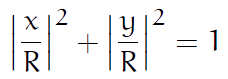
With R the radius of the hair.
We also subtract a circular cylinder of radius r, with r < R,
representing the center of the hair (the medulla) which has a weak birefringence.
Thus, the birefringent thickness e of the sample, crossed by a reflecting light is :
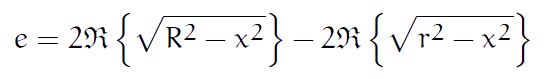
Using these two equations, we can simulate images of Kmax for each
pairs of hair thickness and birefringence, in order to deduce the birefringence
of this hair by comparing the simulation with the actual Kmax measurement.
All these simulated images are compiled in an abacus (Fig. 4) :
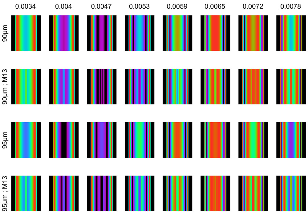
Figure 4 : Hair (partial) birefringence chart with diameters of 90 and 95 µm.
the symbol M13 designates the thickness model with a central medulla, of radius r = 13/100 R.
References :
[1] D. H. Goldstein, Polarized Light, Revised and Expanded. CRC Press, Jun. 2003.
[2] Lattouf, R.et al, "Picrosirius Red Staining: A Useful Tool to Appraise Collagen Networks in
Normal and Pathological Tissues", Journal of Histochemistry & Cytochemistry, 62(10), 751–758 (2014).
[3] Peno-Mazzarino, L. et al., « The K-probe® imaging system as a new tool to analyze human skin
aging », COMET 2019.
Our customers testify
It could help the development of cosmetic assets
Xavier GAILLARD